Figure 8.
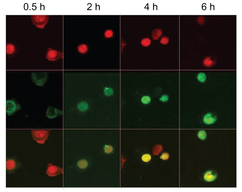
Intracellular distribution of fluorescein isothiocyanate-labeled DNA plasmid/chitosan/polyethylenimine/peptide complexes was observed with a confocal fluorescence microscope in HepG2 cells at 0.5, 2, 4, and 6 hours postincubation.
Notes: The upper panel shows the nucleus (red) and the middle panel shows the fluorescein isothiocyanate plasmid (green). The lower panel shows the overlay of the top two panels.

