
Fig. 6.
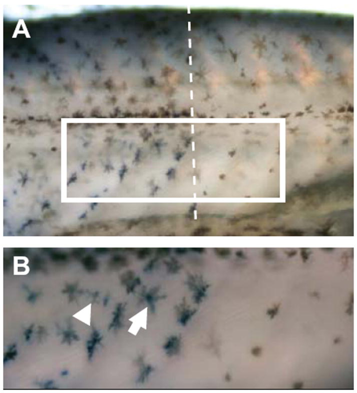
Whole mount in situ hybridization of Pmel in freshwater sticklebacks. Expression of Pmel on the flank of a freshwater stickleback is visualized as blue staining, while pigmented melanophores appear brown. (A) Pmel expression is higher in dark bars (left side of dashed line) than in light bars (right side of dashed line). (B) Enlarged view of the boxed region in (A) showing that Pmel is expressed in pigmented melanophores (arrow) as well as within unpigmented cells that are likely to be undifferentiated melanophores (arrowhead).