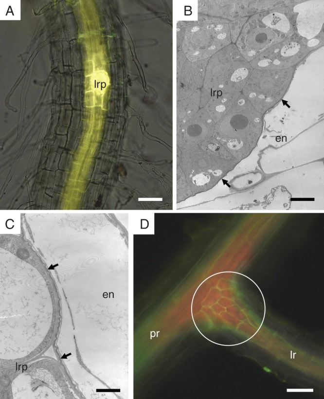
Fig. 6.

The deposition of suberin lamellae related to the development of the lateral root. (A) Endodermal cells surrounding the lateral root primordium are the first endodermal cells depositing suberin lamellae on their primary cell walls (fluorescent microscopy of a whole-mount root cleared and stained by Fluorol Yellow 088). (B and C) Lateral root primordium is covered by endodermal cells containing a protective layer of suberin lamellae (arrows); observed by TEM. (D) A collet of short cells (circle) develops at the base of the lateral root and interconnects the endodermal network of the primary root with the network of a newly formed lateral roots (fluorescent microscopy of whole-mount root cleared and stained by Fluorol Yellow 088). Arrow, suberin lamellae; circle, region of endodermal collet; en, endodermis; lr, lateral root; lrp, primordium of lateral root; pr, primary root. Scale bars (A) = 50 µm; (B) = 5 µm; (C) = 1 µm; (D) = 80 µm.
