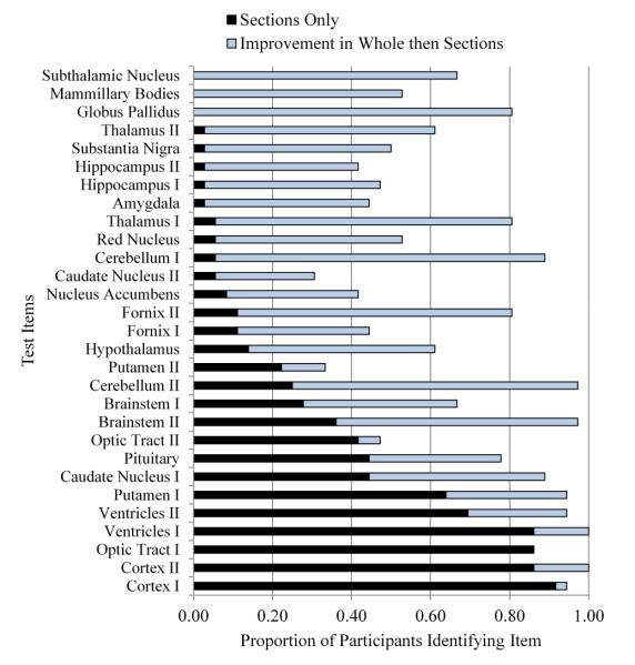
Figure 3.
Proportion of participants correctly identifying neuroanatomical structures in Trial 1 of sectional anatomy learning, broken down by structure, depth of section, and experimental condition. All images were in the coronal (or frontward) orientation. Depth of section is indicated by the roman numerals I and II. Structures are ordered by performance in the sections only group. [Color figure can be viewed in the online issue which is available at wileyonlinelibrary.com]

