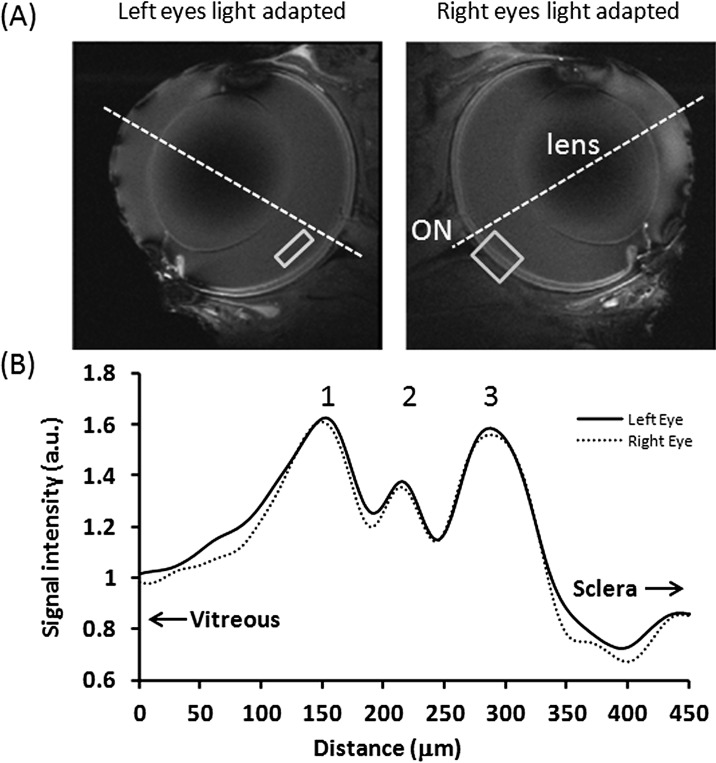
Figure 1. .
(A) MEMRI. (B) Intensity profiles across the retinal thickness of two light-adapted eyes from the same animals at 20 × 20 × 700 μm. Normalization was applied with respect to the vitreous. The vitreous ROI shows the typical region used for normalization. The retina ROI shows the typical region where intensity profile is obtained. The dotted lines approximate the posterior pole.

