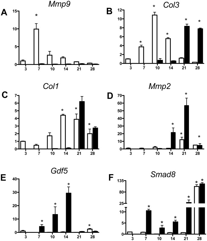
Figure 1. Early expression of neo-tendon associated genes during flexor tendon healing in Mmp9.
−/− mice. Gene expression of (A) Mmp9 (B) Col3a1, (C) Col1a1, (D) Mmp2, (E) Gdf5, and (F) Smad8 in FDL tendon repair tissue over time up to 28 days post-op. Total RNA was extracted and pooled from five tendon repairs per time-point and processed for real-time RT-PCR. Gene expression was standardized with the internal β-actin control and then normalized by the level of expression in day three WT FDL tendon repairs. Data presented as the mean fold induction (over WT day three repairs) ± SEM. * p<0.05 vs. WT day three tendon repair. White bars represent WT mice. Black bars represent Mmp9−/− mice.

