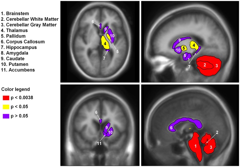
Figure 1. Regional subcortical brain volumes by significance level.
Regions were segmented in Freesurfer. Left and right volumes were averaged and are shown on the right side of the brain only. The brainstem, cerebellar gray matter and cerebellar white matter were significantly reduced in the WFS compared to controls and survived Bonferroni multiple comparison correction (in red; p<.0038). In addition, the thalamus and pallidum were also reduced in WFS compared to controls, but did not survive correction (p<.05, in yellow). Finally, the corpus callosum, hippocampus, amygdala, caudate, putamen and accumbens were not different between groups (p>.05, in purple).

