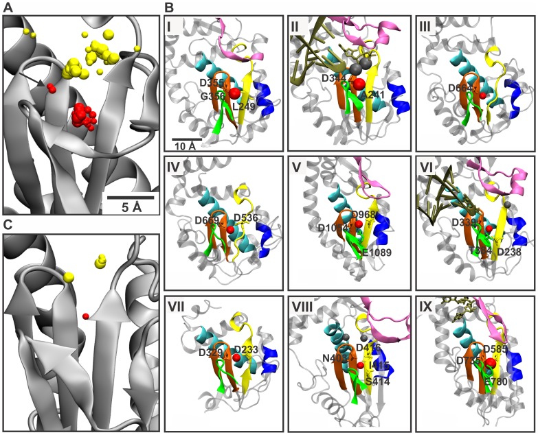
Figure 2. Non-catalytic and catalytic ions in vRdRps.
(A) Structural alignment of vRdRps with bound cations. The catalytic site of the φ6 RdRp (PDBid: 1HHS) polypeptide is depicted with cations from all vRdRp structures. The catalytic ions are colored yellow, and the proposed non-catalytic ions are depicted as red (A and C). The size of the spheres depends on the type of the metal ion ligand. The arrow indicates the position of the non-catalytic ion in the enterobacteria phage Qβ RdRp structures. (B) Magnified views of the catalytic site from selected viral RdRp structures with bound divalent cations. The vRdRp motifs are colored as in Figure 1. Template nucleic acids and nucleotides are shown in tan. Non-catalytic ions are shown as red spheres, and catalytic ions are shown as gray spheres. The amino acids involved in the coordination of the bound non-catalytic ion are indicated. RdRp of (I) rabbit hemorrhagic virus with a Lu2+ ion (PDBid: 1KHV); (II) Norwalk virus with three Mn2+ ions (PDBid: 3H5Y); (III) Dengue virus with a Mg2+ ion (PDBid: 2J7U); (IV) West Nile virus with a Ca2+ ion (PDBid: 2HCN); (V) enterobacteria phage Qβ with a Ca2+ ion (PDBid: 3AGP); (VI) foot-and-mouth disease virus with three Mg2+ ions (PDBid: 2E9T); (VII) poliovirus with a Ca2+ ion (PDBid: 1RDR); (VIII) infectious bursal disease virus with three Mg2+ ions (PDBid: 2R72); and (IX) mammalian orthoreovirus 3 with a Mn2+ ion (PDBid: 1MWH). (C) The catalytic site of the HIV RT structure 1N6Q with cations from all HIV RT structures. The molecules were visualized using program VMD 1.8.7 [30].

