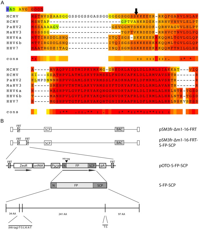
Figure 1. Construction of SCP fusion proteins.
(A) Alignment of beta-herpesvirus SCP sequences with T-Coffee [62]. A naturally occurring glycin-serin-linker like sequence separates the N-terminus in MCMV. (B) Detailed representation of the S-GFP-SCP fusion protein as well as the basic genetic layout of the used mutant viruses carrying a fluorescent protein (FP) and a hemagglutinin tag (HA). GFP or mCherry were used as FPs. The FP could be removed by a simple SpeI-mediated digest resulting in a construct encoding for just a HA-tagged SCP. N marks the duplicated N-terminal region of SCP. The plasmids carrying the fusion constructs were inserted into the MCMV BAC by Flp-mediated recombination.

