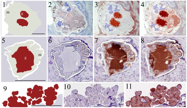
Figure 3. Immunolocalization of SMSs and IgG in scabies mite-infested human skin.
Schematic diagrams 1 and 5 outline the features visible in serial histological sections through a scabies mite in infested human skin. Red staining indicates binding of antibody to protein. Section 2 and 6 probed with pre-immune mouse serum as a negative control remained unstained, while equivalent regions were detected in section 4 and 8 when probed with anti-human IgG, a marker for mite gut [9], and in section 3 and 7 when probed with antibodies against SMSB4 and SMSB3, respectively. Schematic diagrams outline the features visible in serial histological sections through human epidermis containing scabies mite feces 9. Section 10 probed with pre-immune mouse serum as a negative control remained unstained, while equivalent regions were detected in section 11 when probed with anti-SMSB3 and in section 12 with anti-IgG. Staining of feces was also seen in equivalent experiments performed with antibodies against SMSB4 (not shown). Immunohistological co-localization of SMSs and host IgG indicated that both serpins are localized in the mite gut and mite feces within the burrows. b, burrow; f, feces; g, gut; m, mite. Scale bars (100 µM) indicate the magnification level.

