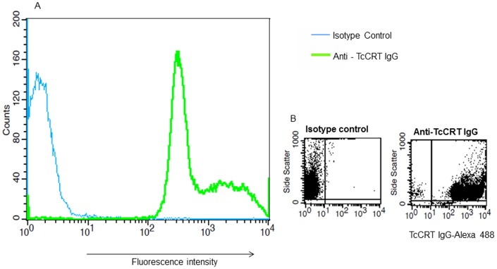
Figure 3. TcCRT is expressed on the surface of T. cruzi trypomastigotes. A.
, Alexa labeled TcCRT IgG binds to the surface of T. cruzi trypomastigotes as revealed by flow cytometry. Shift in the peak of fluorescence indicates that parasite surface is stained with labeled TcCRT IgG. B, Fluorescence intensity of parasites stained with labeled isotype control (left panel) and anti-TcCRT IgG (right panel) showing the high percentage (99.2%) of stained parasites. This figure shows the results of a representative experiment selected from three independent experiments with similar results.

