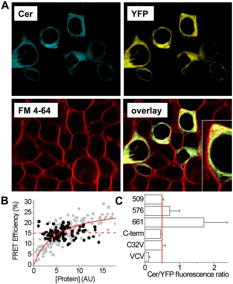
Figure 6. Characterization of 2-color SERCA.
A. Confocal microscopy showed 2-color SERCA was localized in internal perinuclear membranes. Plasma membranes were counterstained with FM 4-64. Inset shows an enlarged view. B. 2-color SERCA showed concentration-independent intramolecular FRET (black points, dotted line). A hyperbolic dependence on protein expression was observed for intermolecular FRET from Cer-SERCA to YFP-PLB (grey points, solid line). C. The brightness ratio Cer/YFP sensitivity ratio for microscopy setup was 0.5 (red line). Deviation from this value suggests a Cer/YFP stoichiometry other than 1∶1. C32V and VCV are controls with 1∶1 and 1∶2 stoichiometry, respectively (25).

