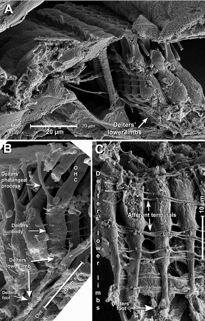
Figure 6.
A: SEM image showing a region of the organ of Corti at the mid region of the cochlear spiral. Note that 1st row DCs’ lower limbs bend slighty before reaching the BM. Scale bar = 20 µm. B: At more apical areas of the mouse cochlea 1st row DCs were more slanted, and the lower limbs bent near 90 degrees away the tunnel of Corti. In this picture the scale bar (= 20 µm) is placed on the BM and parallel to it, making clear that the DC’s leg and the BM have the same orientation. C: Front view of the lower limbs of 1st row DCs at the apical half of the cochlear spiral oriented near parallel to the BM. The feet are fractured and not clearly visible in this picture. Note afferent fibers running on the back and front of the DCs’ lower limbs. Scale bar = 10 µm.

