
Fig. 1.
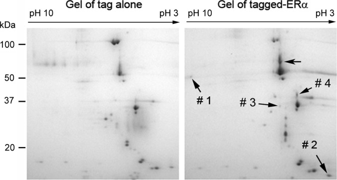
Two-dimensional gel fractionation of the affinity-purified proteins. The tagged ERα and its associated proteins were purified with IgG beads and fractionated with 2-DE. The fractionations of the proteins eluted from control (left panel) and tagged ERα samples (right panel) are shown. LC-MS/MS analysis revealed that spot #1: HADHB; spot #2: calmodulin; spot #3: EFHD1; spot #4: isoform 2 of tropomyosin alpha-3 chain and isoform 1 of tropomyosin alpha-4 chain (Table I).