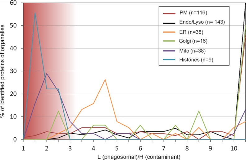
Fig. 2.
SILAC experiment to identify potential contaminations to the phagosome. RAW264.7 macrophages were grown in light DMEM, and phagocytosis was induced for 30 min. These cells were mixed with an equal number of cells grown in heavy labeled DMEM and lysed. Phagosomes were isolated and analyzed by quantitative MS. Subcellular localization of proteins was obtained from Uniprot, and organelles were plotted according to their ratio of light (L) to heavy (H) in bins of 0.5. A light to heavy ratio of ∼1 indicates a potential source of contamination such as mitochondria and histones, whereas most proteins associated to other organelles appear to be genuine phagosome components.

