Figure 6.
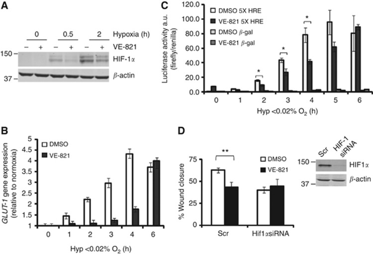
VE-821 inhibits HIF-1 signalling. (A) RKO cells were exposed to ⩽0.02% O2 for the times indicated in the presence of DMSO or 1 μℳ VE-821. Western blotting was carried out for HIF-1α and β-actin (loading control). Representative blots of n=3 experiments are shown. (B) The levels of GLUT-1 mRNA were determined by qRT–PCR. 18S was used as the control. (C) RKO cells were transiently transfected with a HIF-1 reporter construct and exposed to hypoxia as indicated. The levels of luciferase relative to the renilla control are shown. (D) MDA-MB-231 cells were transfected with either Scr (scramble) or HIF-1 siRNA and scratch wound assays were carried out in the presence of DMSO or 1 μℳ VE-821. Western blot showing efficiency of knockdown is shown in inset. The graph represents the percentage of wound closure after 18 h exposure to hypoxia (⩽0.02% O2). Significance values: *P<0.05, **P<0.01.

