Figure 1.
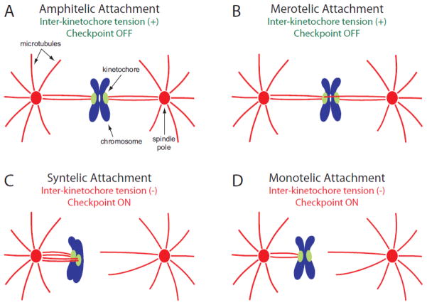
Types of kinetochore attachment to the mitotic spindle. Chromosomes are shown in blue, with associated kinetochores in green. Spindle poles are shown as red ovals, from which microtubules emanate outwards, shown as red lines. (A) At metaphase, kinetochores normally form amphitelic attachments on the spindle. In this orientation, one kinetochore attaches to microtubules emanating from one spindle pole, and its sister kinetochore attaches exclusively to microtubules from the opposite spindle pole. Amphitelic sister kinetochores are bioriented and under tension, and do not activate the spindle assembly checkpoint. (B) A merotelic attachment occurs when a single kinetochore of a sister pair attaches to microtubules emanating from both spindle poles. Although incorrect, these attachments can generate normal levels of tension and do not activate the spindle checkpoint. (C) Syntelic attachments are a form of erroneous linkage in which both kinetochores of a sister pair attach to microtubules emanating from the same spindle pole. Syntelic kinetochores are not under tension and activate the spindle checkpoint. (D) Monotelic attachment describes the case in which one kinetochore attaches to microtubules emanating from one spindle pole, while its sister kinetochore remains unattached. Monotelic kinetochores are not under tension, and the presence of an unattached kinetochore activates the spindle checkpoint. This type of attachment is typically observed early in mitosis, during initial capture of kinetochores by the mitotic spindle.

