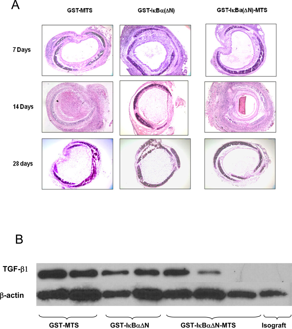
Figure 6.
Infiltration of inflammatory cells in the lumen of the trachea (A). Hematoxylin & Eosin staining reveals the nuclei of inflammatory cells and lymphocytic infiltrations. The lumen of the trachea in the experimental groups injected with GST-IκBα(ΔN)-MTS are less occluded and show less inflammatory cells. Re-epithelialization is more evident in the GST-IκBα(ΔN)-MTS trachea versus the GST-MTS and GST-IκBα(ΔN) tracheas at day 14. (B). Western blot measuring TGF- β in trachea. TGF- β plays a key role in the immune response that culminates in the production of collagen, scarring, and ultimately fibrosis. Without TGF- β acting as part of the inflammatory cascade, fibrosis will not occur. TGF- β levels were significantly lower in the mice that received the NF-κB inhibitor.

