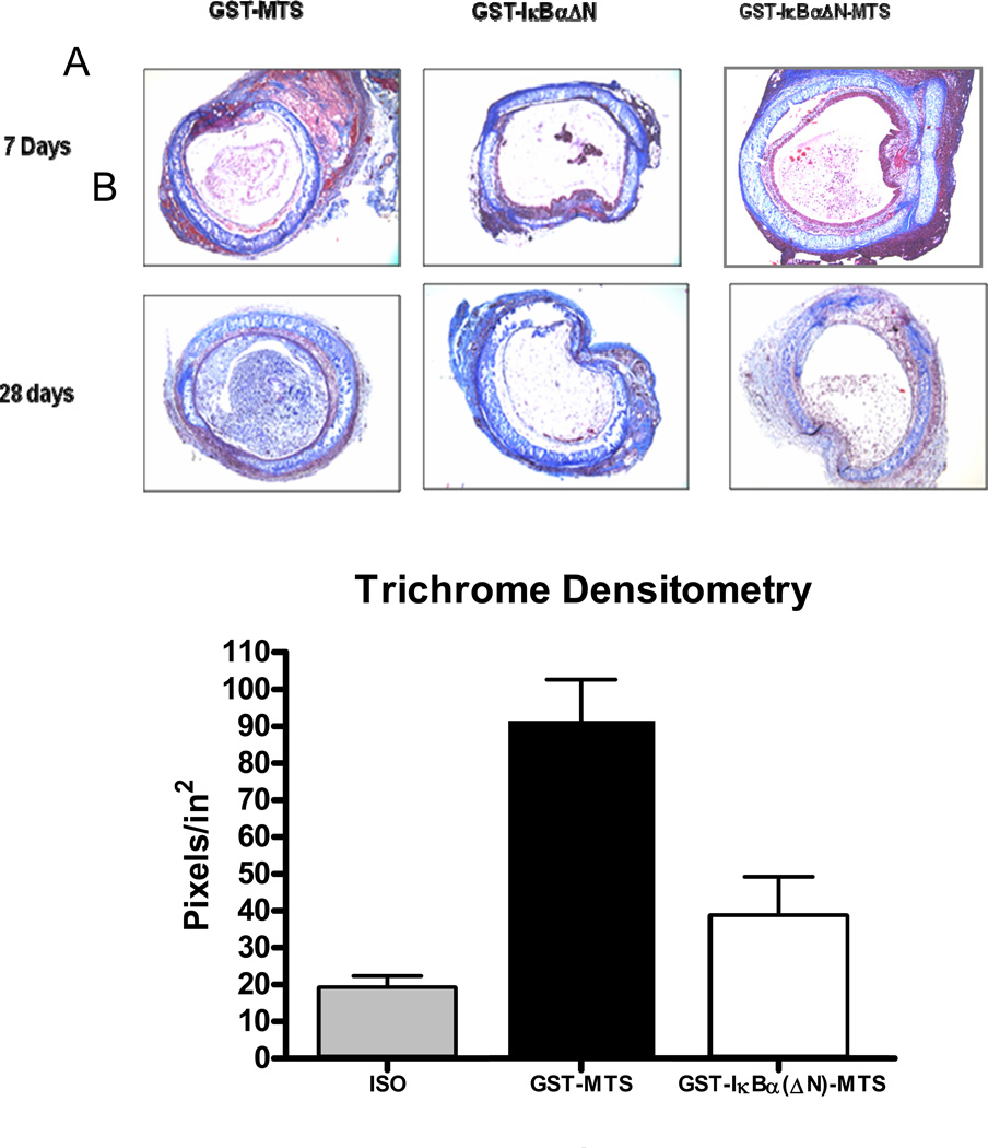
Figure 7.
Collagen deposition in the tracheal lumen. (A). Masson Trichrome staining indicates collagen production as a blue color. While the trachea in both the acute and chronic models show collagen deposition, the GST-MTS and GST-IκBα(ΔN) mice in the chronic model shows much greater intra-luminal collagen deposition than the GST-IκBα(ΔN)-MTS mice. (B). Densitometry quantifies the color blue in the lumen of each photomicrograph

