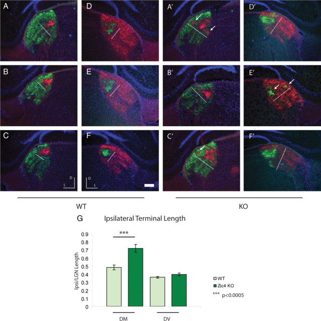
Figure 5.
Ipsilateral projections are disrupted in the rostral LGd of Zic4 KOs. Representative samples of intraocular 488- and 594-CTB tracings to the LGd in control (left, A–C; right, D–F) and Zic4 KO (left, A′–C′; right, D′–F′). Dotted white line, Length of ipsilateral zone along the dorsomedial axis; white arrows, multiple ipsilateral terminals in KO; D, dorsal, L, lateral. Scale bar, 300 μm. G, Ipsilateral terminal length along the dorsomedial (DM) and dorsoventral (DV) axes, expressed as a fraction of the total LGd terminal length.

