Figure 2.
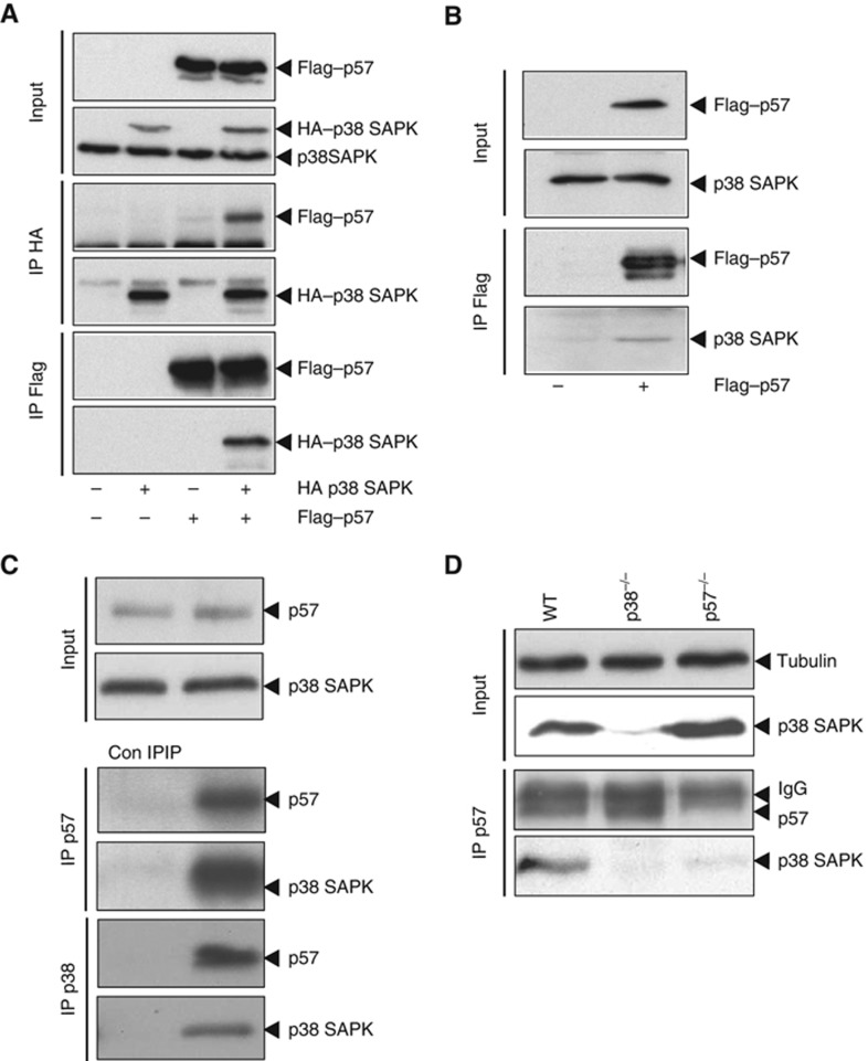
p38 SAPK and p57Kip2 form a stable complex in vivo. (A) HA–p38 and Flag–p57 were transfected into HeLa cells for 48 h. Cell lysates were then immunoprecipitated with either anti-Flag agarose beads or anti-HA coupled sepharose beads and analysed by western blot with anti-HA and anti-Flag antibodies. (B) Flag–p57 was transfected into HeLa cells for 48 h. Cell lysates were then immunoprecipitated with anti-Flag agarose beads and analysed by western blot with anti-p38 and anti-Flag antibodies. (C) HeLa cell extracts were immunoprecipitated with a control IgG (Con IP), anti-p57 or anti-p38 coupled sepharose beads and analysed by western blot with anti-p38 and anti-p57 antibodies. (D) Wild-type, p38−/− and p57−/− MEF cell lysates were immunoprecipiated with mouse anti-p57 coupled sepharose beads and analysed by western blot with anti-p38 and rabbit anti-p57 antibodies. Tubulin was used to monitor the input protein levels. Representative western blots are shown.

