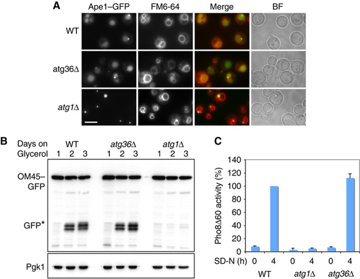
Figure 2.
Atg36 is not required for the Cvt pathway, mitophagy or non-specific autophagy. (A) Cvt pathway activity was assessed by endogenous expression of Ape1–GFP in WT, atg36Δ and atg1Δ cells. Cells were grown for 18 h in glucose medium and vacuoles were stained with FM4-64. Accumulation of GFP in the vacuole indicates normal function of pathway. Bar, 5 μm. (B) Mitophagy of WT, atg36Δ and atg1Δ cells was assayed by western blot analysis of OM45–GFP after culturing cells for up to 3 days in glycerol medium. Samples were taken at 24 h intervals. (C) WT, atg36Δ and atg1Δ cells were assayed for non-specific autophagy by alkaline phosphatase assay. Cells were grown in YPD and shifted to SD-N medium for 4 h. Samples were collected and processed for Pho8Δ60 activity. The results represent the mean and s.d. of three experiments. WT 4 h starvation is set at 100%.

