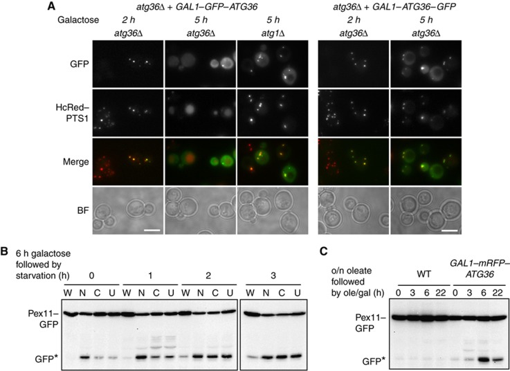
Figure 3.
Atg36 localises to peroxisomes and induces their autophagic degradation. (A) N- (left) and C- (right) GFP-tagged ATG36 under control of the GAL1 promoter was induced in atg36Δ or atg1Δ cells. Early (2 h) and later (5 h) time points after induction are shown. Pexophagy is assessed using the peroxisomal marker HcRed–PTS1 expressed from the constitutive HIS3 promoter. (B) Pex11–GFP western blot showing pexophagy in WT cells grown 6 h in galactose medium (0) then switched to starvation medium for 1, 2 or 3 h as indicated. (W), WT (empty plasmid); (N), GAL1–mRFP–ATG36; (C), GAL1–ATG36–mRFP; (U), GAL1–ATG36. WT shows pexophagy by endogenous Atg36. (C) Pex11–GFP pexophagy of WT cells containing either empty plasmid (WT) or GAL1–mRFP–ATG36 grown 18 h in oleate medium (0) then switched to oleate medium containing 2% galactose and harvested at 3, 6 or 22 h as indicated.

