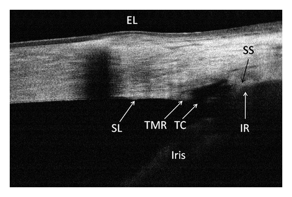
Figure 6.

A cross-sectional OCT image of the nasal angle following trabectome surgery. This frame-averaged image shows that the posterior trabecular meshwork has been removed, leaving a 374 μm wide trabecular cleft (TC) and an anterior trabecular meshwork remnant (TMR). Although scleral shadowing has caused the iris root (IR) to appear indistinct, it was possible to trace its position by contiguity with the iris. The limbal girdle of Vogt is the cause of the shadowing in the peripheral cornea. (Courtesy of Brian A. Francis, MD, USA), reprinted with permission from SLACK Incorporated: Huang D, Duker JS, Fujimoto JG, Lumbroso B, Schuman JS, Weinreb RN. Imaging the Eye from Front to Back with RTVue Fourier-Domain Optical Coherence Tomography. Thorofare, NJ: SLACK Incorporated; 2010.
