
Figure 4.
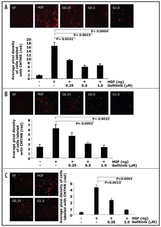
Inhibition of EGFR by gefitinib blocked HGF-mediated cell invasion. (A) NMuMG cells, (B) PyVmT and (C) MDA-MB-231 cells were labeled with a red fluorochrome and plated in the upper chamber, the lower chamber contained either serum free media, HGF or HGF + gefitinib (0.25 μM, 0.5 μM or 1.0 μM) for 8–12 h. Cells that migrated to the underside of the matrigel coated filter were fixed and mounted on slides. Images of at least four fields per filter were taken at 20X at constant exposure. The number of migrated cells were quantified by assaying the pixel density of the labeled migrated cells using software from Scion Image and Adobe Photoshop. Results are presented as average of pixel density of at least four fields ± standard error of the mean of at least four replicates.