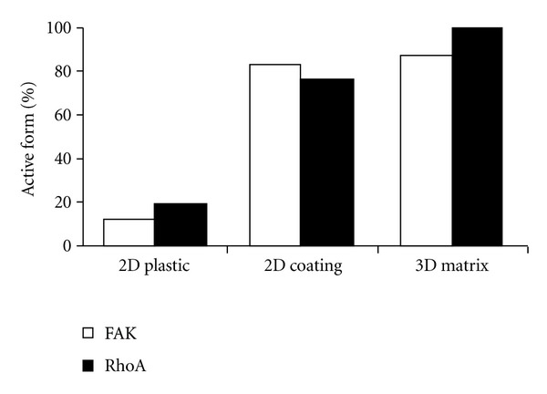
Figure 2.

Type I collagen protects HT1080 cells from doxorubicin-induced dephosphorylation of FAK and RhoA. After 24 h of exposure to doxorubicin (5 and 10 nM), cells cultured on plastic or 2D coated type I collagen were directly lyzed, except for those cultured inside 3D matrices that were beforehand digested by collagenase P. The expression and the activation state of FAK and RhoA were quantified by western blot. Y-axis corresponds to the percentage ratio of active form of FAK or RhoA in doxorubicin-treated cells with respect to untreated cells.
