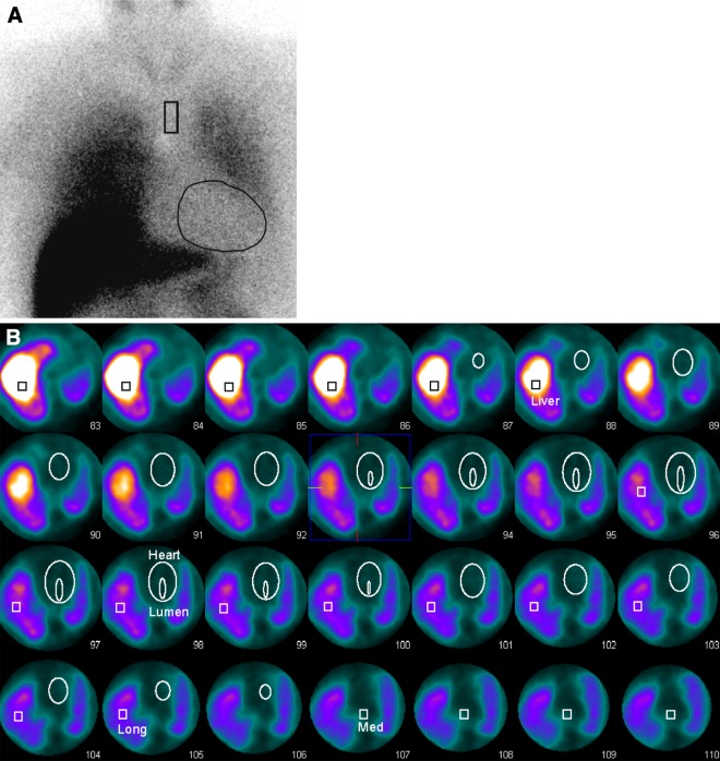
Figure 2.
I-123 MIBG acquisition of a patient from the NDD population (age 60 years). This patient suffers from Parkinson’s disease and diabetes mellitus, but has no history of cardiac diseases. A The early planar image shows a severely reduced accumulation of I-123 MIBG in the cardiac region, but intense uptake in the liver and lung. (eHM = 1.21, dHM = 1.15, WOR = 0.42). B The VOIs for the left ventricle, lumen, mediastinum, right lung and liver are drawn into the SPECT acquisition. Based on these VOIs indices for the HM (eHM = 1.09, dHM = 0.91, WOR = 3.75), MM (eMM = 0.41, dMM = 0.26, WOR = −0.31) and ML (eML = 0.60, dML = 0.52, WOR = 0.08) are determined

