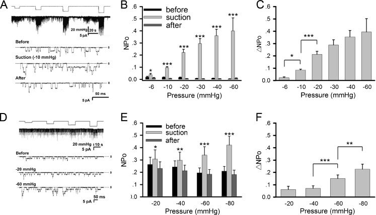
Fig. 1.
Mechanosensitivity of AChRs in muscle cells. AChR activity was studied in cultured Xenopus myotomal muscle cells (a–c) and C2C12 myotubes (d–f). a Sample traces of ACh-induced single-channel currents from Xenopus muscle cells recorded with the patch-clamp method under negative pipette pressure of different magnitudes. Pipette ACh concentration = 0.2 μM, holding potential = +70 mV, cell-attached mode. b Channel activity NPo and c its difference ΔNPo under different negative pressures. For the NPo plot, values before (black), during (gray), and after (dark gray) negative pressure application are shown. Data are mean ± SEM, number of patches n = 9, 91, 46, 46, 44, 18 for negative pressures of −6, −10, −20, −30, −40, −60 mmHg, respectively. d AChR single channel currents recorded from C2C12 myotubes under different negative pressures. ACh concentration = 0.5 μM. e NPo values before (black), during (gray), and after (dark gray) negative pressure application. f ΔNPo data from C2C12 cells. Data from 20 patches at each negative pressure were pooled. Statistics: *p < 0.05; **p < 0.01; ***p < 0.001 (Student’s paired t test). For NPo plots, comparisons were made between values obtained during and before negative pressure application

