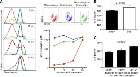
Figure 5. Functional response of the cultured peritoneal macrophages.
(A) Activation (phosphorylation) of p38 kinase in response to stimulation with LPS. Representative histograms of macrophages (red) and eosinophils (blue) that were cultured together but subsequently analyzed with or without first gating out eosinophils (green). Quantification of p38 activation upon stimulation with LPS. The data represent MFI. (B) IL-6 production by cultured peritoneal cells with or without eosinophils. The data represent a mean ± sem. Comparison was performed using Student's unpaired two-tailed t test. (C) IL-6 production by the macrophages cultured with eosinophils in a dose-dependent fashion. The data are mean ± sem.

