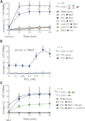
Figure 2. Sublytic concentrations of PVL prime human PMNs for fMLF-stimulated production of O2−.
(A) Priming for release of O2−. PMNs (1×10⁶) were incubated with 1 nM PVL (LukF-PV+LukS-PV), 1 nM iPVL, 1 nM individual PVL subunits (LukF-PV or LukS-PV), or 100 ng/ml LPS for 30 min and then activated with 1 μM fMLF for 20 min, as indicated. Alternatively, PMNs were incubated first with fMLF for 30 min, followed by PVL (as indicated). Results are the mean ± se of three to six separate experiments. *P < 0.05 for the indicated comparisons using a one-way ANOVA and Tukey's post-test. Also, the difference between PVL-primed (closed blue circles) or LPS-primed (open black triangles) PMNs was significant at 5 min. (B) Concentration-dependent, PVL-mediated priming of PMNs for enhanced production of O2−. PMNs were incubated with the indicated concentrations of PVL for 30 min and then activated ± fMLF for 20 min. Results are the mean ± se of five separate experiments. *P < 0.05 versus 0 min using a one-way ANOVA and Dunnett's post-test. (C) Anti (α)-LukS-PV antibody inhibits PVL-mediated PMN priming. PMNs were incubated with 1 nM PVL ± 10 μg/ml anti-LukS-PV or rabbit IgG (Rb IgG) for 30 min and then activated ± 1 μM fMLF for 20 min as indicated. Results are the mean ± se of six separate experiments. *P < 0.05 for the indicated comparisons using a one-way ANOVA and Tukey's post-test. There was also a significant difference between PVL-primed PMNs ± rabbit IgG (open and blue boxes) and PMNs alone (Xs) at 5 min.

