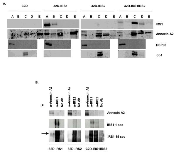
Figure 6. Annexin A2 and IRS1 are expressed in overlapping subcellular compartments and weakly co precipitate in 32D-IRS1 cells.
A. 32D, 32D-IRS1, and 32D-IRS1IRS2 cells were lysed using a subcellular fractionation kit. The subcellular fractions were prepared and analyzed by western blotting with anti-IRS1 and anti-Annexin A2 antibodies. A= cytoplasmic extract, B= membrane extract, C= soluble nuclear extract, D= chromatin bound nuclear extract, E= cytoskeletal extract. The images for 32D cells and 32D-IRS1 cells were derived from the same exposure of the same film. Antibodies to HSP90 and Sp1 were used as positive controls for the cytoplasmic and soluble nuclear fraction, respectively. B. 32D cells expressing IRS1, IRS2, or both were treated with IL-3 for 30 minutes. Cell lysates were prepared and immunoprecipitated with anti-IRS1, anti IRS2, or anti-Annexin A2. Lysates were loaded on a gel and separated by SDS-PAGE. Western blots were probed with anti-Annexin A2 or anti IRS1 antibodies. Several exposures of anti-IRS1 blot are shown. The images for each cell line were derived from the same exposure of the same film.

