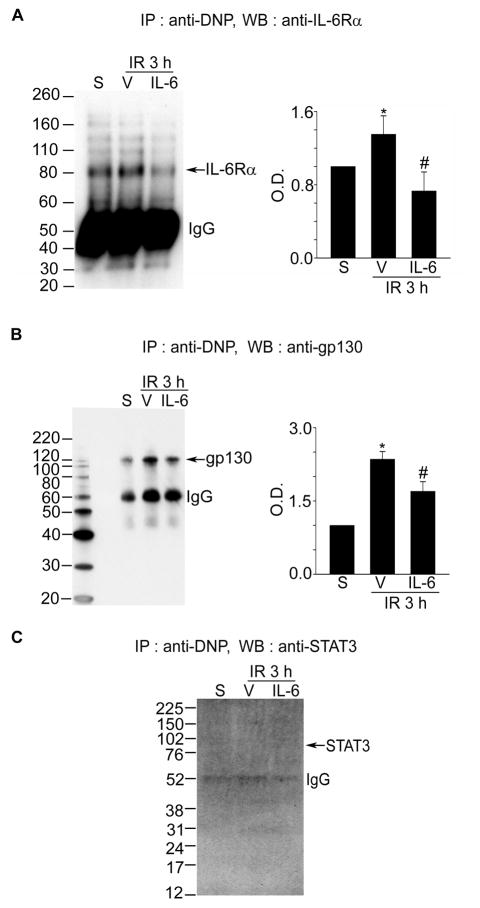
Figure 2.
Oxidation of each component of IL-6R in mouse cerebral ischemic reperfusion. A–C, Co-IP assays for analysis of protein oxidation in the IL-6R components after 3 hours of reperfusion following tFCI with or without IL-6 injection. Summary graphs depicting the band intensity of all Co-IP data. *P<0.05 vs. sham, #P<0.05 vs. vehicle (n=4 per group). IR indicates ischemia reperfusion; S, sham; V, vehicle; IP, immunoprecipitation; DNP, 2,4-dinitrophenyl; WB, Western blot.

