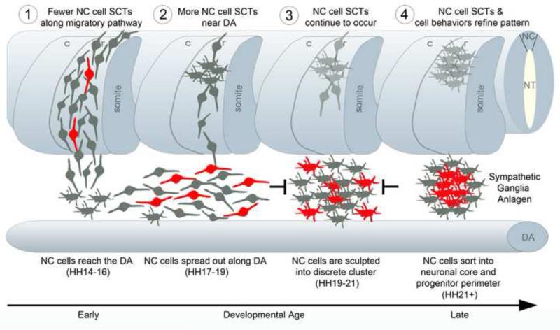
Figure 6. Model of how spontaneous calcium transients refine the substructure of the primary SG anlagen.
Schematic spatiotemporal model of the migration of NC cells forming the SG and the frequency of spontaneous calcium transients (SCT). 1) SCT's occur as NC cells migrate from the NT towards the dorsal aorta (DA) but fewer cells than in later stages exhibit an SCT. 2) NC cells near the DA exhibit SCT's more frequently as they move in an anterior and posterior directions along the DA. 3) NC cells continue to exhibit SCT's as the repulsive forces in the interganglionic regions and attractive forces between NC cells increase. 4) Cells with SCT's promote migration to the center of the cluster forming the neuronal core. Those cells without SCT's move more peripherally forming the progenitor perimeter. Cells in red portray an SCT.

