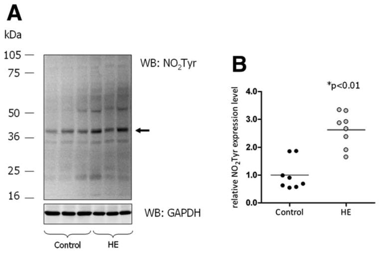
Fig. 1.
PTN in the cerebral cortex from control and HE patients. Protein extracts prepared from the cerebral cortex were analyzed for tyrosine- nitrated proteins by western blot analysis. GAPDH served as a loading control. (A) Western blots from cerebral cortex from three representative control and three HE patients, respectively. The arrow indicates the band used for densitometric quantification. (B) Densitometric quantification of western blot results. Expression of tyrosine- nitrated proteins is given relative to GAPDH expression. Each case included in this study is shown separately in the vertical scatter plot; bars represent the mean values. *Significantly different compared with the control group.

