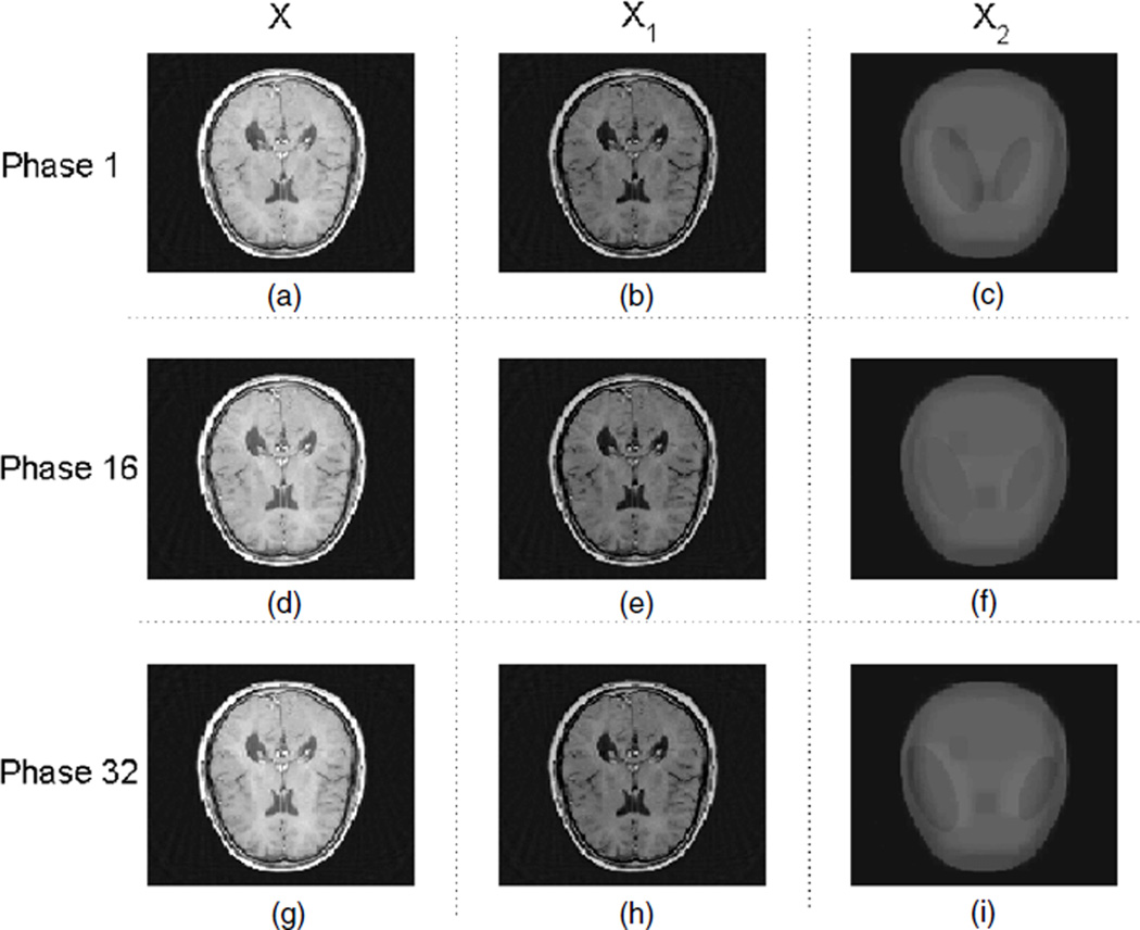
Figure 6.
Reconstructed images with the RPCA-4DCT model for phantom 2; (a), (b) and (c) are the total image X, the low-rank component X1 and the sparse component (in tight frames) X2, respectively, at phase 1, i.e. X = X1 + X2. Similarly, (d), (e) and (f) correspond to X, X1 and X2 at phase 16, and (g), (h) and (i) correspond to X, X1 and X2 at phase 32.

