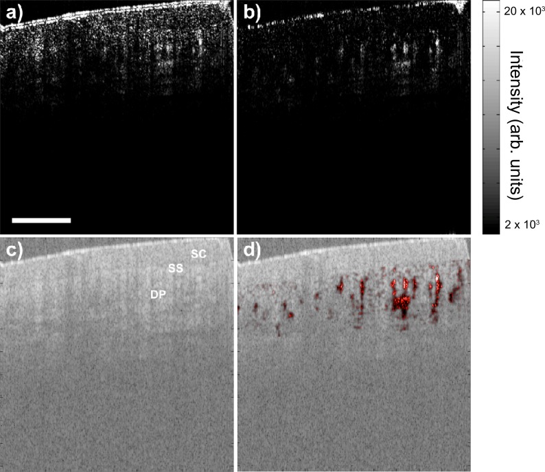
Fig. 3.
B-mode SV image (a) without (Media 1 (2.4MB, AVI) ) and (b) with (Media 2 (2.1MB, AVI) ) subpixel image registration realignment displayed at 54fps during human fingernail fold imaging. (c) Corresponding real-time structural OCT image. (d) Structural image overlaid with SV image (b) in post-processing. Different layers of tissue can be delineated easily in the structural image, while microvasculature can be clearly visualized in the SV image. SC: stratum corneum; SS: stratum spinosum; DP: dermal papillae. Scale bar is 500μm.

