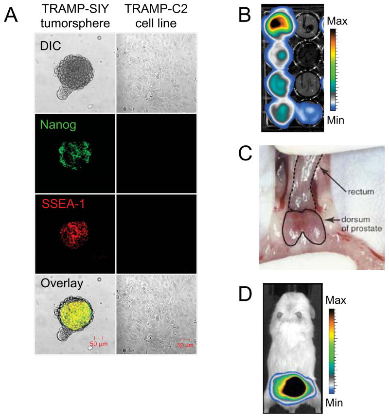
Fig. 5. Orthotopic bioluminescent mouse model of prostate cancer to evaluate the efficacy of adoptive T cell therapy.
(A) Confocal micrographs of a tumor spheroid derived from a primary TRP-SIY tumor (left panel), compared to cells from the TRAMP-C2 cell line (right panel), stained for the stem cell markers Nanog and SSEA-1. Scale bar, 50 μm. (B) Selection of TRP-SIY tumor clones, stably transduced with secreted Gaussia luciferase, by bioluminescent imaging. (C) Orthotopic implantation of Gaussia-luciferase-positive TRP-SIY tumor cells into the dorsal lobes of the mouse prostate. (D) 4 weeks post-implantation, TRP-SIY tumors are visualized by in vivo bioluminescent imaging.

