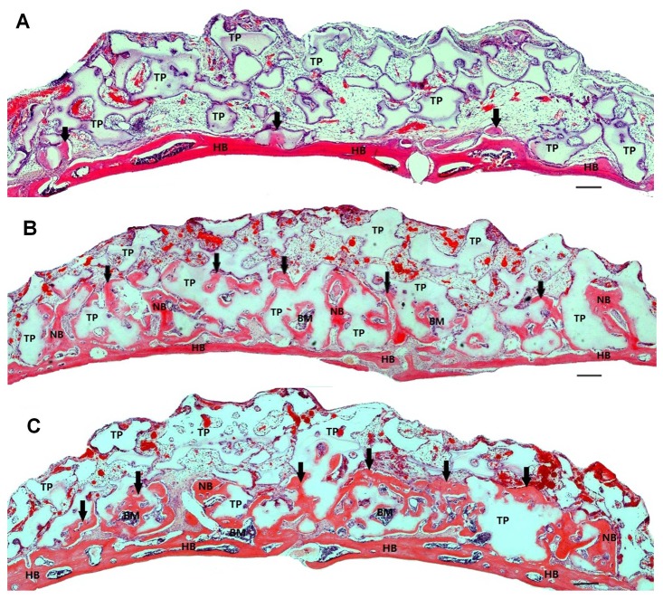
Figure 2. Typical histological micrographs of newly formed bone 4 weeks after transplantation.
Coronal plane sections were stained with hematoxylin and eosin. (A): β-TCP alone induced only a limited amount of new bone formation, and most particles were embedded in loose connective tissue. (B) and (C): A large amount of new bone and immature bone marrow were generated on the calvarial bone of mice transplanted with BMAC (B) or PRP (C). The newly formed bone was sufficiently integrated with the host bone. The TCP particles were in the process of being resorbed and replaced with newly formed bone. The scale bars represent 100 µm. Abbreviations: HB, host bone; NB, new bone, also indicated by arrows; BM, bone marrow; TP, β-TCP particle.

