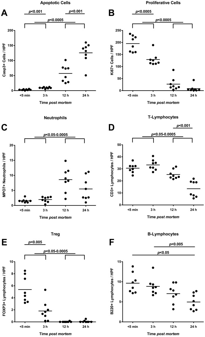
Figure 4. Inflammatory, proliferative and immune cells in ilea of deceased conventional mice.
Mice harboring a conventional intestinal microbiota were sacrificed by cervical dislocation and kept at constant ambient conditions. Ileum biopies were taken at defined time points postmortem as indicated on the x-axis. The average numbers of (A) apoptotic cells (positive for caspase-3, Casp3), (B) proliferative cells (positive for Ki67), (C) neutrophilic granulocytes (Neutrophils, positive for MPO7), (D) T-lymphocytes (positive for CD3), (E) regulatory T-cells (positive for FOXP3, Treg), and (F) B-lymphocytes (positive for B220) from at least six high power fields (HPF, x400 magnification) per animal (n = 8) were determined microscopically in immunohistochemically stained ileal paraffin sections (see Methods). Means (black bars) and levels of significance (P-values) as determined by the Student’s t-test are indicated. Data shown are representative for three independent experiments.

