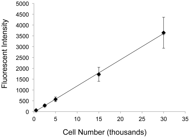
Figure 1. Standard curve of GFP fluorescent signal from lysed GFP-MSCs.
GFP-MSCs were counted using a hemocytometer and set numbers of cells were spun down in a centrifuge. Cell pellets were lysed and solution fluorescence was measured by a fluorometer. The coefficient of determination for the linear regression was 0.999, showing a very strong linear correlation between GFP-MSC number and solution fluorescence.

