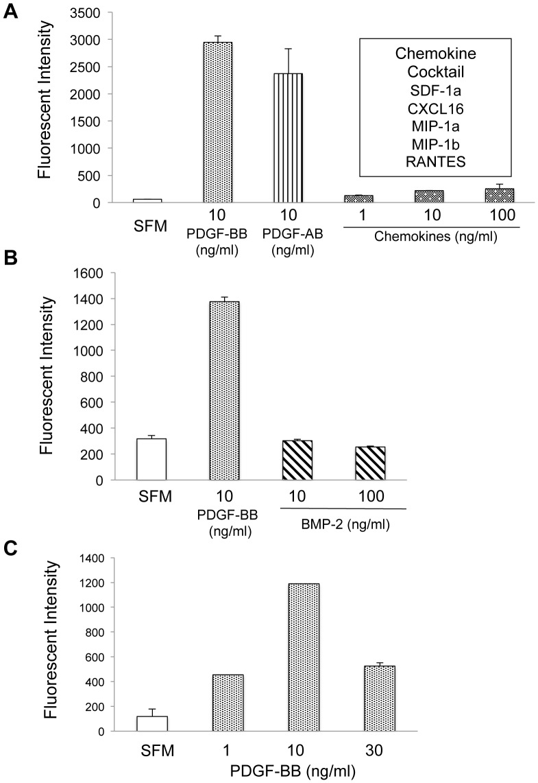
Figure 2. Chemotactic responses of MSCs.
GFP-expressing MSCs (4×10⁴) were seeded onto the top of transwell chambers, with various cytokines/chemokines placed in the bottom of the chambers; some wells contained serum-free media (SFM) as a negative control. After a 20 hr incubation at 37°C, the GFP-MSCs that had migrated across the transwell membrane were lysed and quantitated by measuring fluorescence intensity of GFP. The following chemoattractants were evaluated: A) recombinant human PDGF-BB, PDGF-AB, or a mixture of SDF-1α, CXCL16, MIP-1α, MIP-1β, and RANTES, each at the indicated concentrations (ng/mL) (representative of 3 independent runs) B) PDGF-BB and BMP-2 (representative of 3 independent runs) and C), varying concentrations of PDGF-BB showing dose response.

