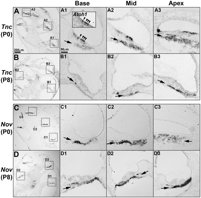
Figure 4. Expression patterns of Tnc and Nov in the cochlea during neonatal development.
Expression patterns of Tnc (A,B) and Nov (C,D) were examined by in situ hybridization at P0 (A,C) and P8 (B,D). (A,B) Tnc transcripts were observed in the basilar membrane in an increasing gradient toward the apex at both P0 and P8 (A1–B3, arrows). Tnc expression was also positive in the differentiating hair cells (Atoh1 expression domain) at P0 (inset in A1, arrowheads), but was down-regulated at P8. (C,D) Nov was expressed in the basilar membrane at P0 and P8. Nov expression levels were relatively constant at P0 along the cochlear duct and gradually decreased toward the apex by P8 (D). Scale bar in A (200 µm) applies to B, C, and D; scale bar in A1 (50 µm) applies to A1–A3, B1–B3, C1–C3, and D1–D3.

