FIGURE 2:
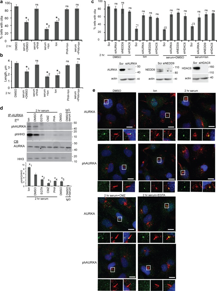
AURKA, NEDD9, and HDAC6 are intermediates in Ca2+/CaM-induced ciliary disassembly. (a) Ciliated hTERT-RPE1 cells were treated for 2 h with the AURKA inhibitor 0.5 μM PHA-680632 (PHA), 500 nM Ion, or DMSO, with or without the addition of serum as indicated, and then percentage of ciliated cells was assessed after 2 h. *1, p = 0.012; *2, p = 0.032; *3, p = 0.013; ns, not significant. (b) Analysis of the length of cilia based on immunofluorescence in A. *1, p = 0.021; *2, p = 0.0091; *3, p = 0.0016; ns, not significant. (c) Top, percentage of ciliated cells 48 h after transfection with siRNA to AURKA, NEDD9, or HDAC6 or with scrambled (Scr) control siRNA, and treatment for 2 h with 500 nM Ion and/or serum. *1, p = 0.0078; *2, p = 0.014; *3, p = 0.0068; ns, not significant. Bottom, depletion of AURKA, NEDD9, and HDAC6 was confirmed by Western blot. For a–c, an average of 200 cells was counted in each of three experiments; error bars, SE. (d) AURKA activity at 2 h after serum treatment of ciliated cells is induced by treatment with 500 nM Ion or inhibited by treatment with PHA (0.5 μM), CMZ (5 μM), or EGTA (0.5 mM), based on assay of immunoprecipitated AURKA autophosphorylation or phosphorylation of histone H3 (HH3, 3 μg). Graph indicates ratio of phHH3 to total HH3, quantified from analysis of three blots; error bars, SE. *1, p = 0.007; *2, p = 0.0023; *3, p = 0.0094; *4, p = 0.0038; *5, p = 0.0072. Immunoglobulin G (IgG) was used as negative control for immunoprecipitation. (e) hTERT-RPE1 cells were treated with the CaM inhibitor CMZ (5 μM), EGTA (0.5 mM), Ion (500 nM), or DMSO for 2 h, with or without serum as indicated. All images are merged panels of acetylated α-tubulin (red), T288-autophosphorylated (active) phAURKA, or total AURKA (green) and DAPI (blue). Scale bar, 10 μm. Right, magnification of indicated boxed structures.

