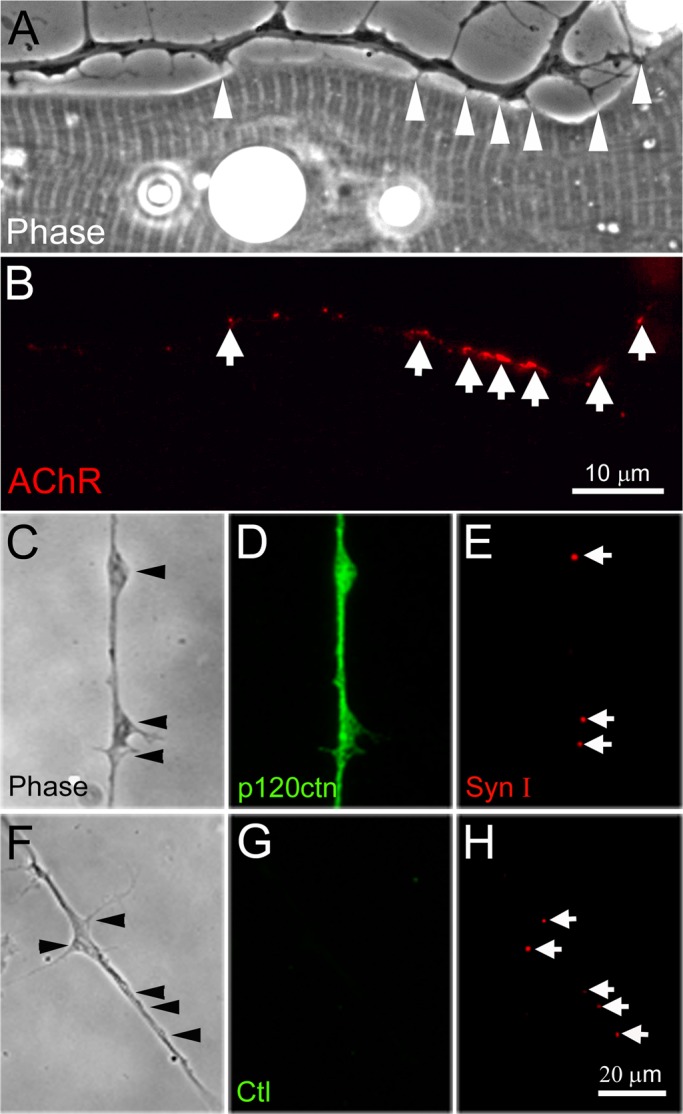
FIGURE 1:

Filopodia and SV clusters in Xenopus spinal neurons. (A, B) Axonal filopodia contacted muscle cells in cocultures (arrowheads in A) and R-BTX labeling showed that AChR clusters formed at these contacts (arrows in B). (C–E) Eighteen-hour-old spinal neurons were fixed and coimmunolabeled with anti-p120ctn and anti–synapsin-I antibodies. A diffuse distribution of p120ctn was observed in axons with no specific concentration at SV puncta. (F–H) No signal was detected if the primary antibody was replaced with anti–hemagglutinin tag antibody (G). SV clusters labeled by anti–synapsin-I antibody often existed at the base of filopodia or at varicosities (C, E and F, H).
