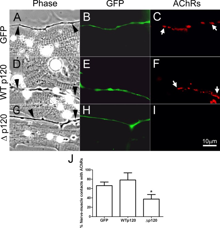
FIGURE 8:
The function of neuronal p120ctn in NMJ formation. Spinal neurons expressing GFP (A–C), WTp120 (D–F), or Rho-mutant Δp120 (G–I) were cocultured with 3-d-old muscle cells cultured from uninjected embryos, and NMJ formation along nerve–muscle contacts was examined after 1 d. The expression of exogenous proteins in spinal neurons was confirmed by GFP fluorescence, and labeling with R-BTX identified AChR clusters in muscle. Neurons expressing GFP (B) and WTp120ctn (E) effectively induced AChR clusters where they contacted muscle cells (C, F), with clusters present at 66.3 ± 7.7 and 78.3 ± 15.3% of contacts between muscle cells and GFP- and WTp120ctn-neurons, respectively (J). In contrast, Δp120ctn-expressing neurons (H) induced AChR clustering (I) at only 37.5 ± 9.7% of contacts with muscle cells (J). Arrowheads point to innervating axons, and arrows mark AChR clusters. Mean and SEM are shown. *p < 0.05, compared with control.

