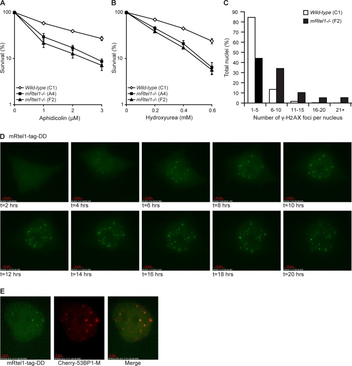
FIGURE 2:
mRtel1 is required during S phase. (A, B) Sensitivity of wild-type (C1) and two mRtel1-deficient (A4 and F2) ESC lines to the indicated doses of aphidicolin (A) and HU (B). Bars, mean percentage values of four experiments with SDs. (C) Analysis of the number of γ-H2AX foci per nucleus in wild-type (C1) and mRtel1-deficient (F2) ESCs. Foci in 1200 wild-type and 750 mRtel1-deficient nuclei in one focal plane were analyzed. (D) Images from a time-lapse movie of mRtel1-tag-DD expressed in mRtel1-deficient (F2) ESCs and cultured in the presence of aphidicolin (2 μM) and shield-1 (1 μM), both added at t = 0. Three image z-stacks (2.5-μm spacing) were acquired every hour. Projections of three z-stacks are depicted. Scale bar, 10 μm. (E) Colocalization of mRtel1-tag-DD and Cherry-m53BP1-M. Cells were cultured in the presence of aphidicolin (2 μM) and shield-1 (1 μM), and images depicted were taken at 17.5 h. Scale bar, 5 μm.

