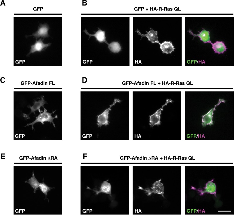
FIGURE 6:
Active R-Ras translocates afadin FL, but not afadin ΔRA, to the cell periphery. Neuro2a cells were transiently transfected with GFP (A), GFP + HA-R-Ras QL (B), GFP-afadin FL (C), GFP-afadin ΔRA + HA-R-Ras QL (D), GFP-afadin ΔRA (E), and GFP-afadin ΔRA + HA-R-Ras QL (F). The cells were fixed at 1 d after transfection. The fluorescence images of GFP (A–F, green in merge) and the immunofluorescence images with anti-HA antibody (B, D, F, magenta in merge) are shown. Scale bar, 25 μm.

