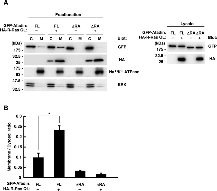
FIGURE 7:
Afadin is localized to the membrane by active R-Ras in a RA domain–dependent manner. (A) Neuro2a cells were transfected with GFP-tagged afadin FL or ΔRA mutant together with HA-tagged R-Ras QL, and the cytosol (C) and the membrane (M) fractions were analyzed by immunoblotting. Na+/K+-ATPase and ERK were used as markers of membrane and cytosol, respectively. Expression levels of the constructs were verified by immunoblotting of the cell lysates (right). (B) The membrane/cytosol ratio of GFP-tagged afadin was analyzed. Data are presented as the means ± SEM of three independent experiments (*p < 0.05, one-way ANOVA with Dunnett's post hoc test).

