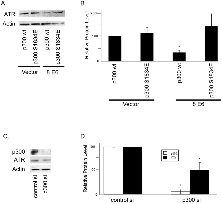
Figure 6. Reduced ATR protein levels in HPV 5 and 8 E6 expressing cells is dependent on p300 degradation.
A. Representative immunoblot showing ATR levels in HFK cells cotransfected with either p300wt or p300 S1834E and vector control or HPV 8 E6. β Actin was used as a loading control. B. Densitometry of immunoblots (n = 3) of ATR protein levels in HPV E6 expressing HFK cells cotransfected with either p300wt or p300 S1834E and vector control or HPV 8 E6. Levels were normalized to β actin for each experiment. Error bars represent standard errors of the mean. * denotes a statistically significant difference from HFK cells cotransfected with vector control and p300wt. C. p300 was knocked down in HFK cells by transfection with a pool of 4 siRNAs targeting p300 or a pool of 4 non-targeting siRNAs. Representative immunoblot showing ATR and p300 levels in these cells HFK cells 72 hours post transfection. β Actin was used as a loading control. D. Densitometry of immunoblots (n = 3) of ATR and p300 protein levels in HFK cells transfected with a pool of 4 p300 targeting siRNAs or a pool of 4 non-targeting siRNAs. Levels were normalized to β actin for each experiment. Error bars represent standard errors of the mean. * denotes a statistically significant difference from HFK cells transfected with control siRNAs.

