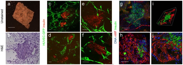
Figure 1. In vitro vascularization of islet multicultures.
10-day-old multi-cellular cultures of mouse pancreatic islets, human ECs and HFFs, grown on 3D PLLA/PLGA polymer scaffolds were analyzed. (a) Whole scaffold, bright field photo; scale bar- 2mm. (b) A paraffin-embedded section stained with H&E; scale bar-200 µm. (c–f) Stereomicroscope confocal images of engineered vascularized pancreatic islet scaffolds stained for insulin (red) and HUVEC-GFP (green). (g–h) Laser scanning confocal image of a cell-embedded scaffold stained for insulin (green), vWF (red) and nuclear content (blue); scale bar-100 µm. (i–j) 3D reconstruction of images collected in (g–h) using the Imaris software.

